WHAT IS HIGH BLOOD PRESSURE?
High blood pressure (HBP) or hypertension means high pressure (tension) in the arteries. Arteries are vessels that carry blood from the pumping heart to all the tissues and organs of the body. High blood pressure does not mean excessive emotional tension, although emotional tension and stress can temporarily increase blood pressure. 

  
Table 1. JNC 7 Blood Pressure Classification
Blood Pressure Classification
Systolic Blood Pressure, mm Hg
Diastolic Blood Pressure, mm Hg
Normal
< 120
< 80
Prehypertension
120-139
80-99
Stage 1 hypertension
140-159
90-99
Stage 2 hypertension
= 160
= 100


    RISK FACTORS


SIGNS AND SYMPTOMS

Most people with high blood pressure have no signs or symptoms, even if blood pressure readings reach dangerously high levels.
Although a few people with early-stage high blood pressure may have dull headaches, dizzy spells or a few more nosebleeds than normal, these signs and symptoms typically don't occur until high blood pressure has reached an advanced — even life-threatening — stage.

COMPLICATIONS 


MANAGEMENT

Blood pressure treatment goals*
140/90 mm Hg or lowerIf you are a healthy adult
130/80 mm Hg or lowerIf you have chronic kidney disease, diabetes or coronary artery disease or are at high risk of coronary artery disease
120/80 mm Hg or lowerIf your heart isn't pumping as well as it should (left ventricular dysfunction or heart failure) or you have severe chronic kidney disease
* Although 120/80 mm Hg or lower is the ideal blood pressure goal, doctors are unsure if you need treatment (medications) to reach that level.
Lifestyle Modifications to Manage Hypertension* (NON PHARMACOLOGICAL)
for more info visit :


INTRODUCTION

imageEpidermoid cysts, sometimes known as sebaceous cysts (a misnomer), contain a soft "cheesy" material composed of keratin, a protein component of skin, hair, and nails.
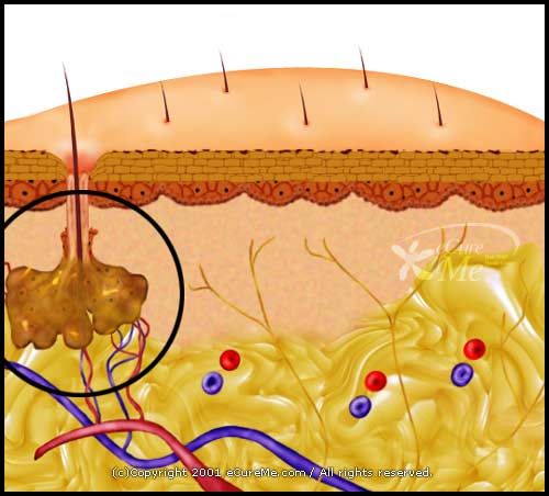
  • Epidermoid cysts form when the top layer of skin (epidermis) grows into the middle layer of the skin (dermis). This may occur due to injury or blocked hair follicles.
  • The lesion may be asymptomatic, but rupture of the epidermoid cyst can result in significant discomfort.


Blocked sebaceous glands, swollen hair follicles, and excessive testosterone production will cause such cysts.


Symptoms


  • Are round cysts or small bumps that are easy to move with your fingers
  • Are usually white or yellow, though people with darker skin may have pigmented cysts
  • Range in size from less than 1/4 inch to nearly 2 inches (a few millimeters to 5 centimeters) in diameter
  • Occur on nearly any part of your body, including your fingernails, but are found most often on your face, trunk and neck

Signs and symptoms of infection, which can occasionally occur, include:
  • A thick, yellow material draining from the cyst that may have a foul odor
  • Redness, swelling and tenderness around the cyst


When to see a doctor

Most epidermoid cysts aren't harmful, but you may want to have them removed for cosmetic reasons. See your doctor if you have a cyst that:
  • Grows rapidly
  • Ruptures
  • Becomes painful
  • Occurs in a spot that's constantly irritated

image

Treatments


  • Inflamed, non-infected cysts may be injected with steroids to reduce inflammation.
  • Incision and drainage can provide immediate reduction in the cyst. However, this is a temporary measure. After this treatment, a cyst will refill with the cheesy contents because the lining of the cyst has not been removed.
  • Cysts may be removed (excised) surgically.


The scalpearsbackface, and upper arm, are common sites for sebaceous cysts, though they may occur anywhere on the body except the palms of the hands and soles of the feet. In males a common place for them to develop is the scrotum and chest. They are more common in hairier areas, where in cases of long duration they could result in hair loss on the skin surface immediately above the cyst. They are smooth to the touch, vary in size, and are generally round in shape.


Non-surgical

Another common and effective method of treatment involves placement of a heating pad directly on the cyst for about fifteen minutes, twice daily, for about 10 days (depending on size and location of the cyst). There is some anecdotal evidence however that this home remedy can lead to infection in a previously uninfected cyst. This may be caused by an over-heated or non-sterile heat pad.

FOR MORE INFO:
http://www.mayoclinic.com/health/sebaceous-cysts/DS00979/METHOD=print
http://www.skinsight.com/adult/epidermoidCystSebaceousCyst.htm
What Is Anemia?
Anemia is a condition that develops when your blood lacks enough healthy red blood cells. These cells are the main transporters of oxygen to organs. If red blood cells are also deficient in hemoglobin, then your body isn't getting enough iron. Symptoms of anemia -- like fatigue -- occur because organs aren't getting enough oxygen.

What Causes Anemia?


What Are the Symptoms of Anemia?


MANAGEMENT OF IRON DEFICIENCY ANEMIA


DIET IN IRON DEFICIENCY ANEMIA

Hemoglobin rich foods 


Hemoglobin rich foods


Nutritional anemia is the most common cause of anemia, of which iron deficiency is the most prevalent nutritional deficiency. In addition to iron, there are other nutrients that prevent or reduce anemia and these include vitamin B6, vitamin B2, vitamin B12, vitamin C, folate and protein. These nutrients are actively involved in the process of blood formation (hemopoiesis) and are termed as hemopoietic nutrients.


Tarsal bones mnemonic
"Traverse City (is) Noted (for) MIchigan's Lovely Cherries": Talus Calcaneus (new row) Navicular (new row) Medial Intermediate cuneiforms Lateral cuneiform Cuboid


FEMUR


TIBIA AND FIBULA
                                              fibula


Anteromedial view of right fibula.


Tibia

Tibia. Anterior (A) and posterior (B) views of the right tibia.


FOOT


HIP JOINT (made of femur head and acetabulum of pelvis)

KNEE JOINT (made of femur, tibia, fibula and patella)


F

ANKLE JOINT (made of tibia, fibula and talus)
File:Ankle.PNG


STEPS :


  1. 1
    Identify your patient                                                        .


  2. 2
    Assemble your supplies.                                            


  3. 3
    Wear gloves                                                                         .


  4. 4
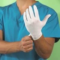
    Decide which arm you will be drawing from or let your patient decide and tie the tourniquet 2-3 inches from the antecubital fossa.              


  5. 5
    If no veins are palpable, have the patient make a fist. Don't allow the patient to pump his/her fist                                                                             .


  6. 6
    Once a suitable vein is found, disinfect the area with the alcohol wipe.                              


  7. 7
    Remove the tourniquet until ready to draw. The specimen will be inaccurate and damage to the patient can be caused if the tourniquet is left on longer than a minute.


  8. 8
    Allow the alcohol on the skin to dry completely.


  9. 9
    Do not touch the venipuncture site again.


  10. 10
    With the needle attached to the tube holder and a tube loosely in the holder, ensure that the bevel of the needle is pointing up.


  11. 11


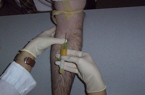
    Anchor the vein from below the intended puncture 
    site using the thumb of the opposite hand, and insert the needle at no more than 15 degrees.                                                          


  12. 12
    Push the tube onto the needle and check for blood flow.


  13. 13
    Allow the tube to fill. Remove the filled tube from the tube holder.                                                    


  14. 14
    Remove the tourniquet once the first tube starts to fill.


  15. 15
    Mix the contents if tube used has additives.


  16. 16
    Fill remaining tubes and repeat previous step. When last tube is filled, remove vacutube.


  17. 17
    Have a piece of gauze ready in the opposite (anchoring) hand, remove needle, and place gauze on top of the venipuncture site. Apply pressure.


  18. 18
    Activate the needle's safety feature and discard the needle in a sharps container.


  19. 19
    Label the tubes.                                                           


  20. 20
    Ensure that the patient has stopped bleeding, and apply tape and gauze, or a bandage to the venipuncture site.


  21. 21
    Discard waste and put materials away              
                                                                                                              .
  22. VIDEO :